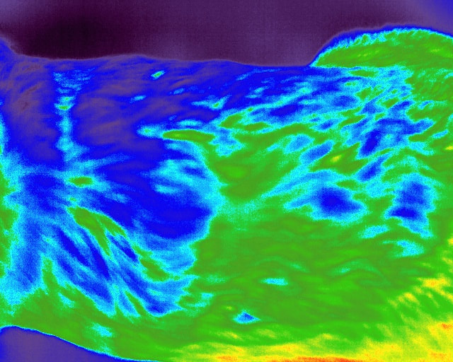
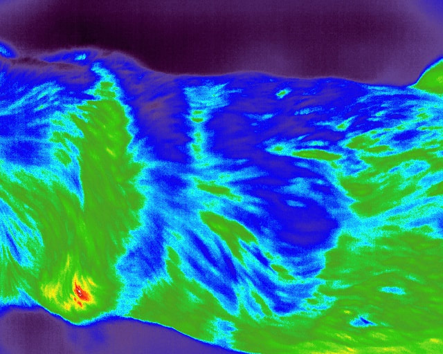
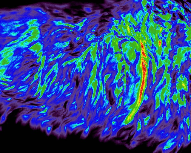
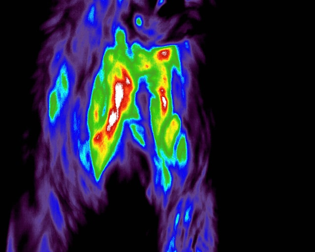
Digital thermography evaluation is included in all orthopedic, rehab, and spinal manipulation therapies performed in a temperature controlled environment.
See pain in a different light - digital thermal imaging using the advanced, top of the line Digatherm Digital Thermal Imaging unit!
The first part of managing pain is appropriately diagnosing it. The way JRIVS starts this process in dogs, cats, and horses is by using digital thermography.
There are dozens of thermography units out there, but there is only one Digatherm! The IR Tablet 640 is designed for the highest resolution to see clearer, using 327,680 points of temperature. This allows for the most reference points for patient symmetry to allow the animal to show us what hurts! This is the real deal, and it is no cell phone app!
Digital thermography compiles and analyzes the radiated energy coming from a patient using infrared light. There is no radiation, therefore no harm will come to the patient!
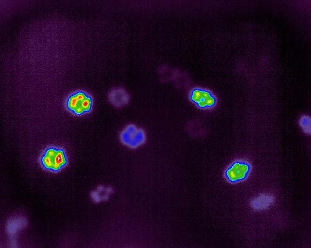
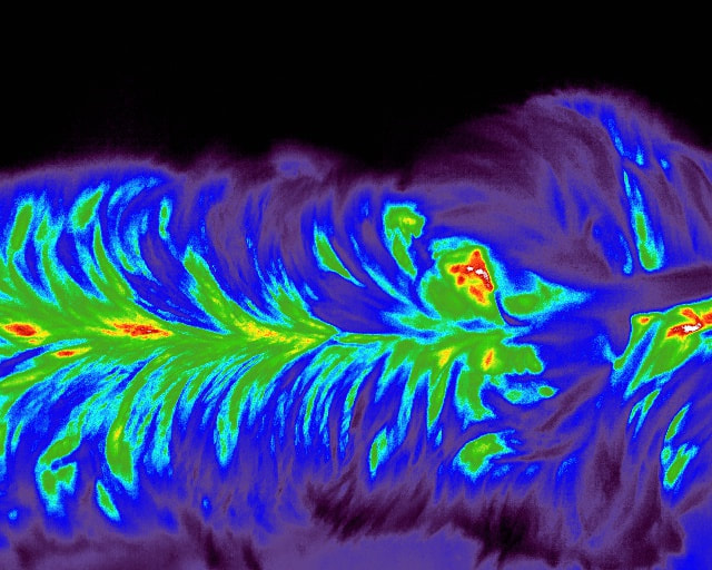
Different levels of radiated energy are interpreted by the digital thermal imaging device and represented as different colors or shades of grey on the computer monitor. This image is called a thermogram. The range and intensity of colors in the thermogram is called a thermal gradient.
A thermogram provides a physiological picture of the area being examined. Structure and function, trauma, and acute and chronic conditions will all change the circulatory activity of the area examined, and produce changes in the thermogram. Not only can these changes illustrate inflammation, but they also illustrate increases or decreases in nerve function. Thermograms not only determine what areas need closer examination, but they also provide a quantitative measurement for functional improvement.
